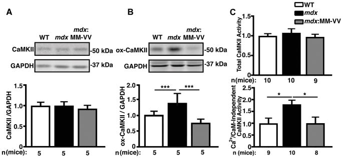
Figure 1. Elevated ox-CaMKII increases autonomously activated CaMKII in mdx mice.
(A) Representative Western blots and quantification of total CaMKII expression level in mdx mouse ventricular lysates compared to WT and mdx:MM-VV. (B) Representative Western blots and quantification of oxidation at M281/M282 (ox-CaMKII) demonstrating significantly increased expression of oxidized CaMKII in mdx mouse ventricular lysates compared to WT and mdx:MM-VV. (C) Fold change in total CaMKII activity induced by Ca2+/CaM (top), and Ca2+/CaM-independent CaMKII activity (bottom). *P<0.05, ***P<0.001.

