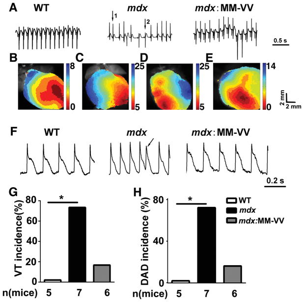
Figure 5. Genetic inhibition of ox-CaMKII attenuates ventricular arrhythmias in isolated mdx heart.
(A) Representative electrogram traces in ex vivo hearts from WT, mdx and mdx:MM-VV mice. Following sinus rhythm (arrow 1), ventricular tachycardia (VT) beats emerged spontaneously (arrow 2) in the mdx mouse heart. (B) Normal activation map of WT mouse heart. (C) Activation maps of mdx mouse heart corresponding to normal beat (arrow 1) and (D) the ventricular tachycardia beat (arrow 2). (E) Representative activation map of heart from mdx: MM-VV mice showing normal activation map. (F) Representative action potential (AP) tracings from WT, mdx, and mdx:MM-VV hearts. Arrow denotes delayed after depolarization (DAD). (G–H) Quantification of VT (G) and DAD (H) incidence in ex vivo WT, mdx, and mdx:MM-VV hearts.

