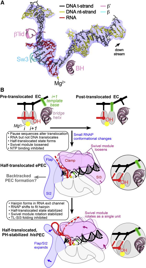
Figure 7. The half-translocated state arises in a PH-minus PEC.
(A) Shown is the 5.5 Å resolution (4.3 Å resolution around the active site and RNA-DNA hybrid) cryo-EM density map (blue mesh) with the superimposed model of the hisPEC-minus-PH nucleic acids. Shown for reference are key RNAP structural elements (Sw3, lid, BH) and the RNAP active-site Mg2+-ion (yellow sphere). Like the hisPEC (Figure 2A), the half-translocated RNA:DNA (shown), but not a pre-translocated or fully translocated RNA:DNA, fit the density map.
(B) A mechanistic model of pausing. The half-translocated state arises in the ePEC when RNAP conformation is loosened, making it susceptible to swiveling (Sekine et al., 2015), and is stabilized by global rearrangement of RNAP including swiveling in the PH-stabilized PEC. Insets depict template DNA and RNA near the active site.
See also Table S3 and Figure S6.

