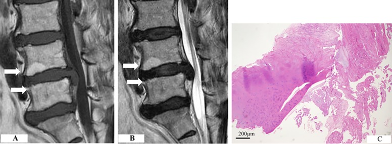
Fig 1. Cartilaginous herniation with Modic type 2 change.
(A) The inferior endplate of L4 and the superior endplate of L5 show increased signal intensity on both T1-weighted scan. (B) The T2-weighted scan showed increased intensity in the same location. (C) Cartilaginous endplates were present in the herniated disc, along with the annulus fibrosus.

