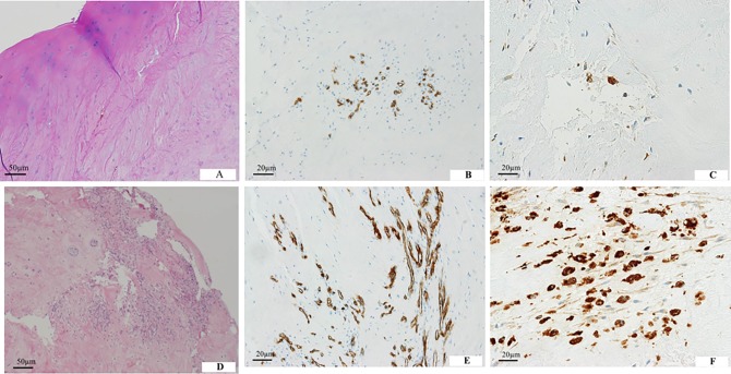
Fig 2. Composition of herniated tissues.
(A) A herniated mass containing hyaline cartilage from the endplate. (B) CD34-positive capillaries are partly observed in the herniated specimen. (C) CD68-positive macrophages are less frequent. (D) Inflamed granulation tissue is observed in herniated specimens without cartilaginous endplates. (E) CD34-positive capillaries are distributed diffusely in the herniated specimen. (F) CD68-positive macrophages are abundant.

