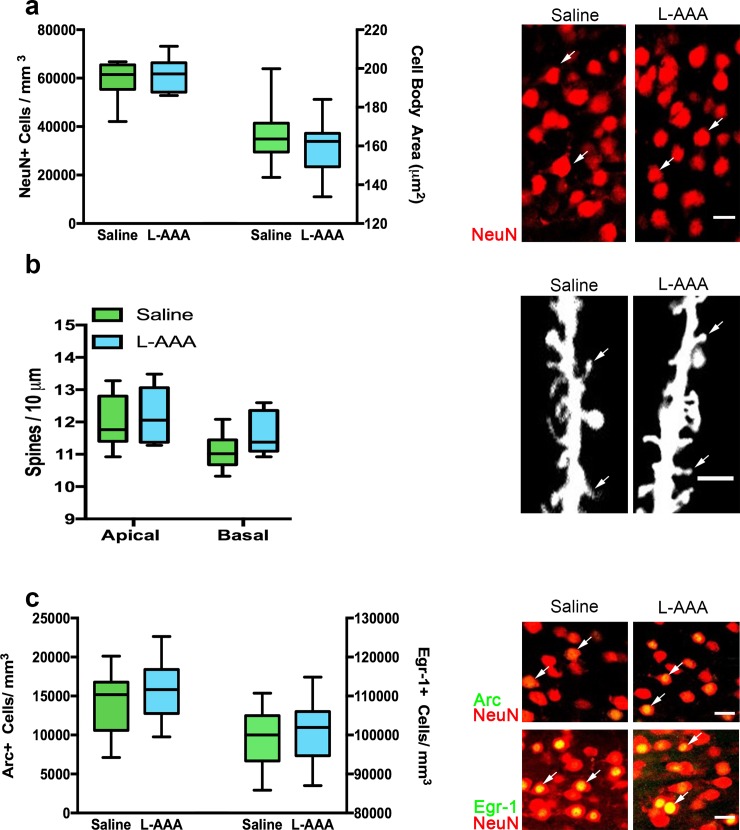
Fig 2. L-AAA does not alter neuron number, size, dendritic spine density or immediate early gene expression in the mPFC.
To rule out the possibility that L-AAA had deleterious effects on neurons, neuronal viability was characterized using several measures. a. Relative to saline-infused controls, both the density and cell body size of NeuN+ layer 2/3 pyramidal neurons was unchanged in L-AAA treated rats (n = 11–12). Confocal images of NeuN+ staining in the mPFC. Scale bar = 20 μm. Arrows point to NeuN stained cell bodies in the mPFC. b. In a separate cohort of rats, no differences in dendritic spine density for both apical and basal secondary and tertiary dendrites of layer 2/3 pyramidal neurons were found when comparing saline and L-AAA treated rats (n = 6). Confocal images of DiI-labeled tertiary apical dendrites in layer 2/3 pyramidal neurons in the mPFC. Scale bar = 5 μm. Arrows point to example spines on along the dendrite. c. No differences in the density of Arc+ and Egr-1+ neurons in layer 2/3 of the mPFC were detected between saline and L-AAA -treated rats (n = 11–12) after completing the ASST. Confocal images of Arc (green) and Egr-1 (green) and NeuN (red) immunolabeling. Scale bar = 20 μm. Arrows point to examples of colabeling between NeuN+ neurons in red and the respective immediate early gene marker in green.

