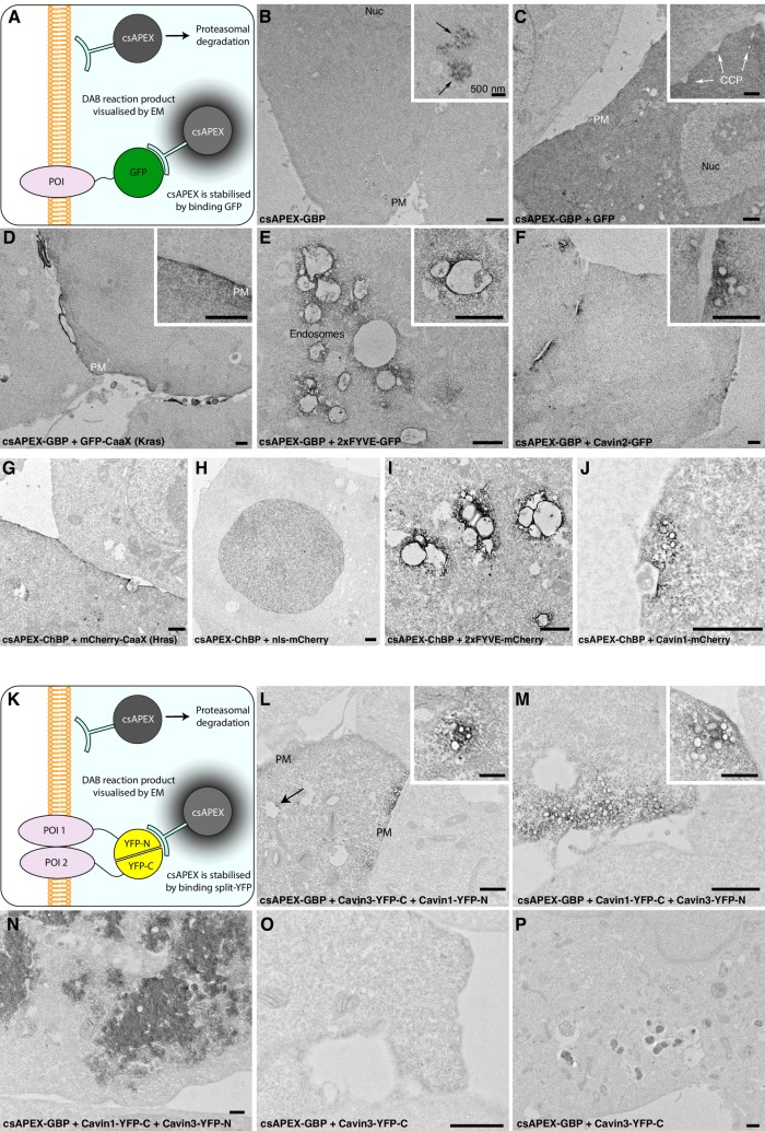
Fig 2. Conditional stabilisation of GBP and ChBP, and detection of protein–protein interactions using bimolecular fluorescence complementation.
A) Schematic illustrating detection of GFP-tagged POIs using csAPEX-GBP. The probe is degraded by the proteasome unless stabilized by interactions with a GFP-tagged protein, resulting in loss of any nonspecific, electron-dense APEX signal when csAPEX-GBP does not bind to its target. B) csAPEX-GBP shows minimal signal when expressed in cells lacking GFP-tagged proteins; only a low level of labelling is detectable in specific regions of a subset of cells (inset, arrows). In contrast, cells co-expressing soluble GFP together with csAPEX-GBP show a strong cytosolic signal (C, quantitated in S2 Fig. A). D-F) Examples of subcompartment-specific labelling in cells expressing GFP-tagged POIs associating with the PM, the early endosomes, and caveolae, respectively. G-H) Examples of subcompartment-specific labelling in cells expressing mCherry-tagged POIs associating with the PM, nucleus, early endosomes, and caveolae, respectively. K-P) Co-transfection of BHK cells with constructs tagged with each half of split YFP along with csAPEX-GBP gives strong and specific labelling at sites of protein–protein interactions. K) Schematic illustrating detection of interactions between two POIs tagged with different halves of a split YFP. csAPEX-GBP is able to bind only when the YFP pair is fully reconstituted and folded. In the absence of a correctly folded GFP derivative, csAPEX-GBP is degraded by the proteasome. L) Cavin1-YFP-N and Cavin3-YFP-C co-expression gives specific labelling associated with PM pits and vesicular profiles characteristic of caveolae. Note the specificity of the labelling, which allows identification of Cavin1/Cavin3 complexes associated with both surface caveolae and putative endocytic caveolar carriers associated with intracellular compartments (arrow). Further examples are shown in S2B and S2C Fig. M) Reciprocal experimental conditions with specific fragments of YFP switched between constructs gives consistent labelling. N) Cells with an abnormally high transfection level show intracellular aggregates of Cavin (compare with caveolar labelling in L and M). O) Control cells transfected with just one split GFP half and csAPEX-GBP show no labelling in the majority of cells. P) APEX positive inclusions are seen in a small percentage of control cells. These are clearly distinguishable from the specific staining of the recombined protein complex (L-M). Further examples are shown in S2 Fig. D. Scale bars: lower magnification = 1 μm; insets = 500 nm. BHK, baby hamster kidney; CCP, clathrin-coated pits; ChBP, mCherry-binding peptide; cs, conditionally stable; GBP, GFP-nanobody/binding peptide; PM, plasma membrane; POI, protein of interest.

