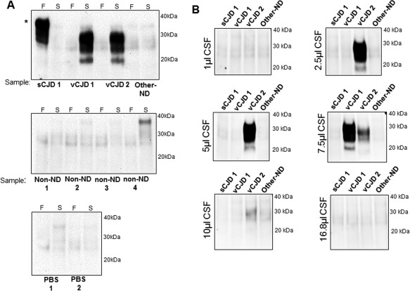
Figure 1.

Detection of vCJD prions in cerebrospinal fluid samples by hsPMCA. (A) CSF samples from one probable and one definite vCJD (vCJD1 and vCJD2, respectively), one sporadic CJD MM1 (sCJD1), one non‐CJD neurodegenerative disease case (Other‐ND), one non‐neurodegenerative case (non‐ND), and two reactions incubated with PBS were evaluated by hsPMCA. Reactions were seeded with a final volume of 8.4 µl of sample. Non‐amplified frozen (‘F’) and amplified (‘S’) samples were analysed. (B) A range of CSF sample volumes were considered for amplification using the same CSF panel. The samples were mixed with an equal volume of substrate (83.2 µl) and normalized to a final 100 µl reaction volume. The samples were subjected to a single round of amplification. Samples were treated with Proteinase K and evaluated by western blotting using 3F4 mAb. Reference molecular mass of electrophoretic markers is shown. (*) Incomplete proteolytic digestion of PrP. [N = 5; 2 vCJD (definite and probable), 1 sCJD, 1 non‐ND, 1 Other‐ND].
