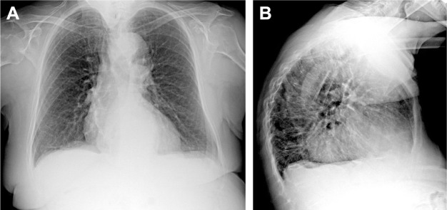
Figure 2.
Chest X-ray in PA (A) and LL (B) projections. Chest X-ray has poor sensitivity to detect COPD; possible findings include prominence of the hilar vessels and decreased peripheral bronchovascular markings, flattened diaphragm due to hyperexpansion and hyperkyphosis and increased lung lucency (especially seen in the retrosternal region in LL projection) and bullae (round focal lucency over 1 cm).
Abbreviations: PA, posterior–anterior; LL, latero-lateral.

