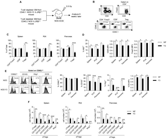
Fig. 6. Selective inability of IL-2RβY3 Tregs to compete with wild-type Tregs in vivo.
(A) Experimental scheme for establishing the mixed bone marrow chimeras. (B) Representative flow cytometry plots of the donor cell populations that were isolated from NOD.SCID recipient mice 8 weeks after transfer. (C) Percentages of the donor cells in the indicated tissues. Data were analyzed by Mann-Whitney test (n=7). (D) Flow cytometric analysis of the expression of Ki67, CD25, and Foxp3 in Tregs from the indicated tissues. Data analyzed by Mann-Whitney test and Wilcoxon signed-rank test for MFIs (n=7). (E) Representative flow plots from the spleen (left) and analysis of the percentage of CD62Llo Tregs and their expression of CD103, Klrg1 and ICOS (right) from the indicated tissues. Data for ICOS (MFI) were analyzed by Wilcoxon signed-rank test; the remaining data were analyzed by Mann-Whitney test (n=7). (F) Analysis of the numbers of the indicated Treg subsets from the indicated tissues based on the expression of CD62L, CD69, CD103, and Klrg1. Data were analyzed by Mann-Whitney test (n=7).

