Fig. 1.

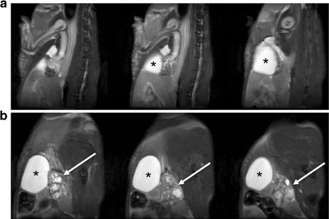
Representative multi-slice T2-weighed MR images of a wild-type control and b Pten/Trp53 mutant mouse with prostate cancer, illustrating locations of enlarged prostate tumor (arrow) and bladder (asterisk).

Representative multi-slice T2-weighed MR images of a wild-type control and b Pten/Trp53 mutant mouse with prostate cancer, illustrating locations of enlarged prostate tumor (arrow) and bladder (asterisk).