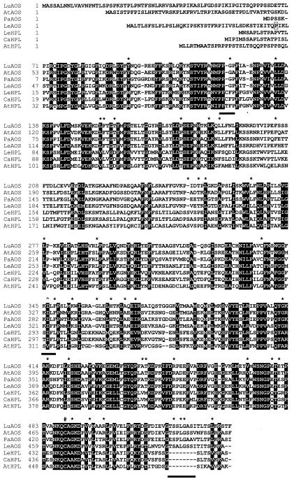
Figure 2.
Comparison of cDNA-deduced protein sequences of plant AOS and HPL genes. LeAOS and LeHPL sequences were aligned, using the ClustalW 1.7 program available at http://mbcr.bcm.tmc.edu/searchlauncher. AOS sequences were from flax (Song et al., 1993; accession no. U00428), guayule (Pan et al., 1995; accession no. X78166), and Arabidopsis (Laudert et al., 1996; accession no. Y12636). HPL sequences were from bell pepper (Matsui et al., 1996; accession no. U51674) and Arabidopsis (Bate et al., 1998; accession no. AF087932). Black boxes indicate amino acid residues that are conserved between all seven CYP74 members. Subfamily-specific substitutions are indicated with an asterisk. The three subfamily-specific motifs discussed in the text are underlined by the black bars. The symbol denotes the T → (I/V) change within the I helix that is a hallmark of CYP74 enzymes. The conserved Cys within the heme-binding domain is marked by a # symbol. The boxed residue (Pro-43) at the N terminus of LeAOS denotes the site where the His-tag was added in the pQE-AOS expression construct.

