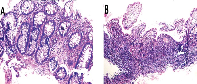
Fig.3.
3A. Backwash ileitis with grade II villous atrophy: Ileal biopsy with surface erosions, villi showing broadening and shortening. The glands showing regenerative changes, the lamina propria infiltrated by mild to moderate chronic inflammatory cells. 3B. Rectal biopsy: mucosal damage, glandular distortion, missing crypts and cryptitis. Lamina propria infiltrated by moderate chronic inflammatory cells. No evident dysplastic or neoplastic changes.

