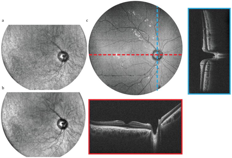
Figure 5.

In vivo SECTR imaging of the posterior retina with >45° (15 mm) FOV in a healthy volunteer (Visualization 1). (a) Raw and (b) 5-frame average of 2560 × 2000 pix. (spectral × lateral) SER images acquired at 200 fps. (c) En face OCT volume projection with representative 5-frame averaged fast- and slow-axis cross-sections (red and blue, respectively). OCT volume was sampled with 2560 × 2000 × 1400 pix. (spectral × lateral × lateral) in 7 s.
