Figure 2.
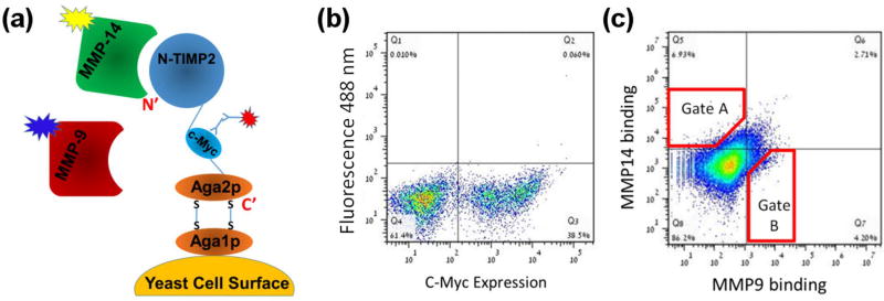
(A) Schematic representation of N-TIMP2 displayed on the yeast surface (adapted from Chao et al.[63]). The N-terminus of N-TIMP2 is free and facing away from the yeast surface. (B) Expression of the construct was monitored by FACS by fluorescently labelled antibodies (red burst in panel A) that bind the c-Myc tag in the construct. (C) Binding of N-TIMP2 to catalytic domains of MMP proteins in solution was monitored by FACS by incubating the N-TIMP2 library with two MMP targets (MMP-9CAT) and (MMP-14CAT) simultaneously and using two different fluorescent labels for each MMP proteins (yellow and blue bursts in panel A). Red polygons represent sample cell collection gates, A for MMP-14CAT and B for MMP-9CAT. The figure here was produced in the initial sorting round on the focused library.

