
Figure 4.
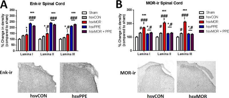
The optical density of Enkephalin (Enk-ir) and co-labeled mu-opioid receptor (mOR-ir) and green fluorescent protein-positive areas of lamina I–III of the dorsal lumbar spinal cord increases after hsvMOR, hsvPPE and hsvMOR + PPE inoculation. A. Quantification of Enk-ir positive areas of the dorsal horn on Day 16 post viral inoculation. There is a significant increase in Enk-ir in hsvPPE and hsvMOR + PPE treatment groups compared to sham and control virus (two-way ANOVA, factors: group × lamina, F(8, 45) = 7.1, P < 0.001, hsvCON). Below: Histology images of Enk+ areas of the dorsal horn of the spinal cord with control virus (left) and hsvPPE (right). B. Quantification of MOR-ir positive areas of the dorsal horn on Day 16 post viral inoculation. Below: Histology images of MOR+ areas of the dorsal horn of the spinal cord with control virus (left) and hsvMOR (right). There is a significant increase in MOR-ir in all lamina of hsvMOR, hsvPPE and hsvMOR + PPE treatment groups compared to sham and control virus (two-way ANOVA, factors: group × lamina, F(8, 45) = 18.7, P < 0.001; hsvCON). # vs. sham; # significant at P<0.05; ## significant at P<0.01; ### significant at P<0.001. * vs. control virus-treated (hsvCON);* significant at P<0.05; ** significant at P<0.01; *** significant at P<0.001. Bonferroni post-hoc for multiple comparisons.