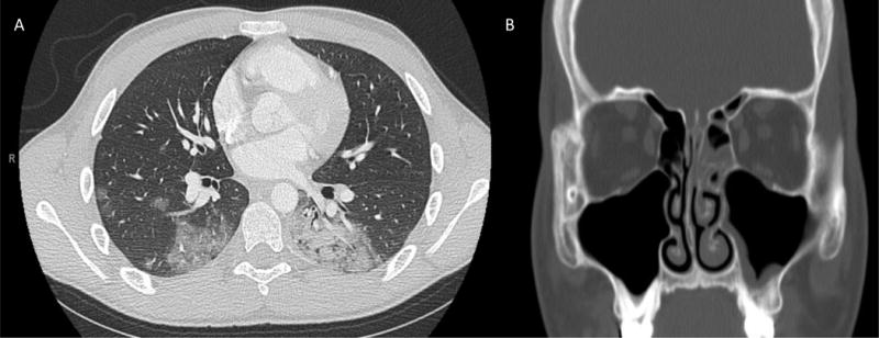
Figure 1.

(A) Chest CT scan chest with contrast at day 5 of induction showing diffuse scattered ground-glass and dense airspace opacities in the right upper and middle lobe and left lower lobe. (B) Sinus CT with contrast showing mild to moderate mucosal thickening of left maxillary and ethmoid sinuses.
