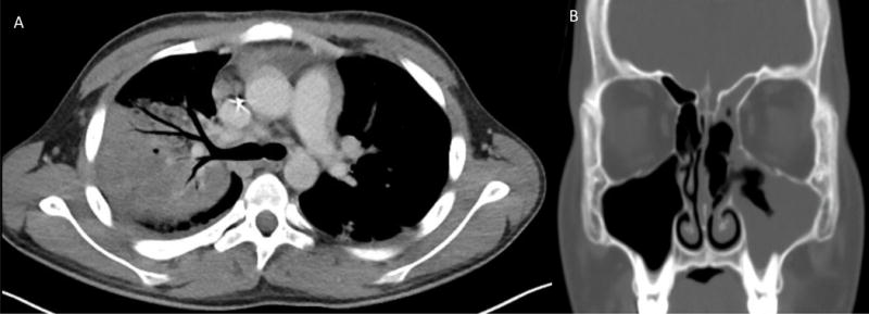
Figure 3.

(A) Chest CT scan with contrast at day 36 after initiation of induction chemotherapy showing right upper lobe consolidation, ground-glass airspace disease in the left lower lobe and right sided pleural effusion. (B) Sinus CT without contrast showing worsened mucosal thickening of left maxillary and ethmoid sinuses.
