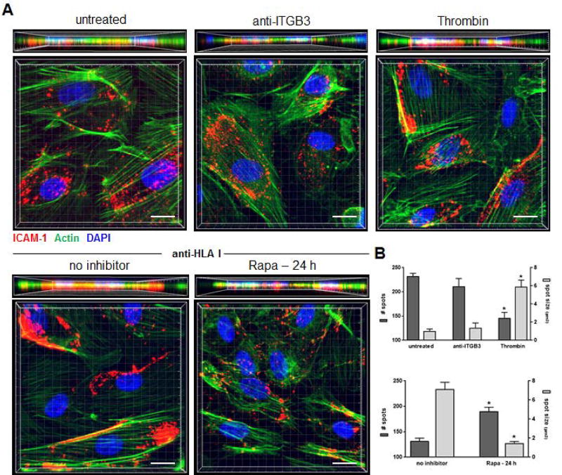
Figure 4. mTOR is required for ICAM-1 clustering in HLA class I Ab-activated ECs.
Primary endothelial cells (ECs) were pre-treated with Rapa or no inhibitor for 24 h, before they were stimulated with Ab against Integrin β3 (anti-ITGB3), HLA I (anti-HLA I; clone W6/32) or thrombin for 5 min, then CFSE-labeled MM6 cells were allowed to adhere for 20 min. ECs were stained by immunofluorescence for ICAM-1 (red), as well as Phalloidin for actin filaments (green) and DAPI to detect cell nuclei (blue). (A) Shown are representative three-dimensional volumetric confocal images of ECs following indicated treatments. scale bars=10 µm. (B) Imaris 3D analysis software was used to quantify ICAM-1 clustering. Shown are the number of individual spots per cell detected in the red channel as well as their size, and results are expressed as the mean number of spots or mean three-dimensional size (µm3) ± SEM of each treated group over untreated ECs (n≥30 cells per group). *P<0.05 by unpaired two-tailed Student’s t test.

