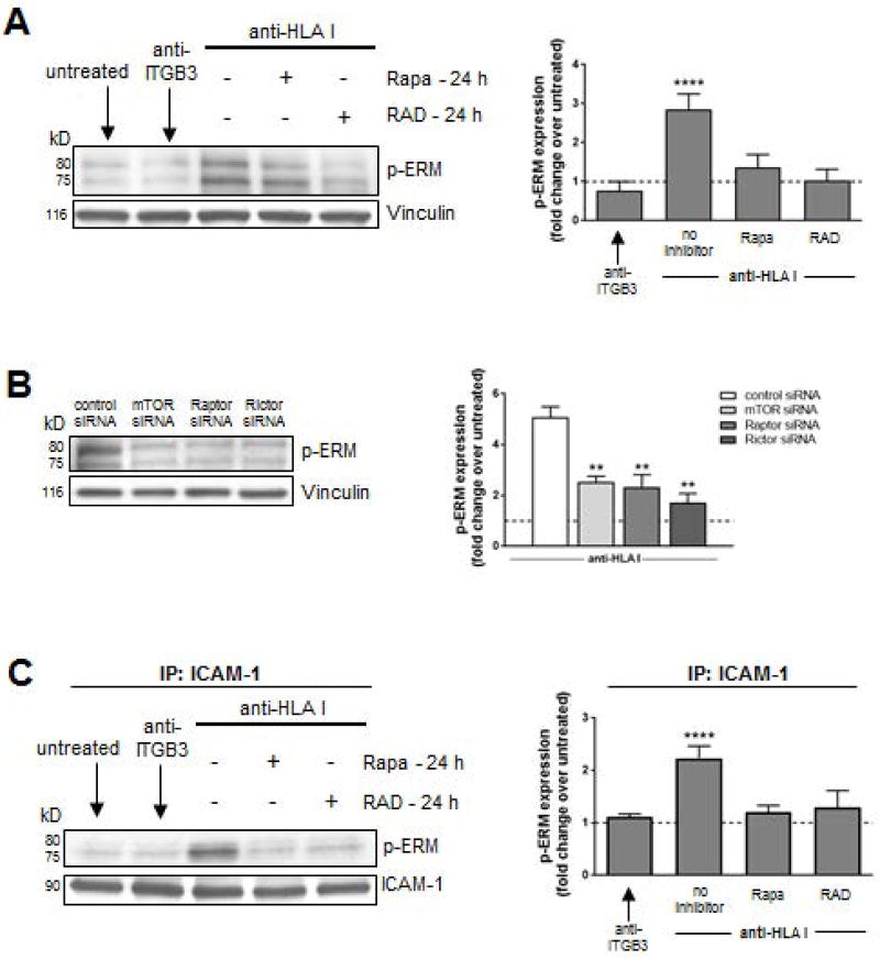
Figure 5. ERM phosphorylation is impaired by mTOR inhibition in HLA class I Ab-activated ECs.
ECs were pre-treated with Rapa or RAD for 24 h and stimulated with HLA I Ab (anti-HLA I; clone W6/32) for 10 min. (A) Cells were lysed and proteins were separated by SDS–PAGE followed by immunoblotting to detect phosphorylated EzrThr456/RadThr564/MoesThr558 (p-ERM) as well as Vinculin to confirm equal loading of proteins and were quantified by densitometry scan analysis using ImageJ. Results are expressed as the mean fold increase in p-ERM expression over untreated ECs ± SEM over 3 independent experiments. (B) ECs were transfected with either control siRNA, or siRNA directed against mTOR, Raptor (MTORC1), or Rictor (MTORC2) before treatment with anti-HLA I for 10 min. Cells were lysed after 48 h and proteins were separated by SDS–PAGE followed by immunoblotting to detect p-ERM as well as anti-Vinculin mAb to confirm equal loading of proteins and were quantified by densitometry scan analysis using ImageJ. Results are expressed as the mean fold increase in p-ERM expression over untreated ECs ± SEM over 3 independent experiments. (C) ECs were pretreated with Rapa or RAD for 24 h and stimulated with anti-HLA I for 10 min. Cells lysates were immunoprecipitated (IP) with anti-ICAM-1 followed by immunoblotting to detect p-ERM as above. Results are expressed as the mean fold increase in p-ERM expression over untreated ECs ± SEM over 3 independent experiments. **P<0.01; ****P<0.0001 by two-way ANOVA with Tukey’s multiple comparisons test.

