Fig. 1.
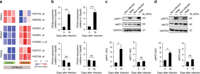
The PI3Kγ signaling is enhanced in the heart tissue of mice after experimental infection with T. cruzi. a Transcriptome analysis for the expression of Pik3ca, Pik3cd, and Pik3cg genes in non-infected C57BL/6J mice or 18 days post infection with T. cruzi. b RT-PCR analysis of the mRNA expression of Pik3ca, Pik3cb, Pik3cd, and Pik3cg genes in the heart tissue of C57BL/6 non-infected mice (n = 7) or 18 days post infection with T. cruzi Y strain (n = 11). Gapdh was used as a housekeeping gene. c Representative western blots and analysis of phosphorylated (p) and total (t) AKT1 expression in the heart tissue of non-infected C57BL/6 mice or 18 days post infection with T. cruzi (n = 6). GAPDH was used as a loading control. d Representative western blots and analysis of phosphorylated (p) and total (t) AKT2 expression in the heart tissue of non-infected C57BL/6 mice or 18 days post infection with T. cruzi (n = 6). GAPDH was used as a loading control. AU refers to arbitrary units (c, d). *P < 0.05, **P < 0.01, ***P < 0.001, and ns = no statistical significance (unpaired Student’s t-test in b–d). Data are representative of two (b–d) independent experiments (mean ± s.e.m in b–d)

