Fig. 4.
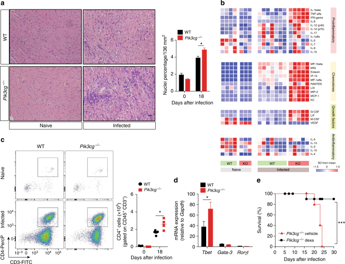
PI3Kγ controls the heart inflammation after infection with T. cruzi. a Representative images from H&E staining of heart tissue from non-infected WT (n = 6) and Pik3cg−/− (n = 6) mice or 18 days after infection with 103 trypomastigote forms of T. cruzi Y strain. Scale bar = 50 μm. b Heat map demonstrating the profile of cytokines and chemokines produced in the heart tissue of non-infected WT and Pik3cg−/− mice or 18 days post infection with 103 trypomastigote forms of T. cruzi Y strain. c Representative flow cytometry plots for the analysis of CD4 staining and quantification of the number of positive cells in the heart tissue of WT (n = 5) and Pik3cg−/− (n = 4) naive mice or infected with T. cruzi Y strain. d PCR analysis of mRNA expression of the transcription factors Tbet, Gata-3, and Rorγt in the heart tissue of WT (n = 4) and Pik3cg−/− (n = 4) mice. Gapdh was used as a housekeeping gene. e Survival rate of Pik3cg−/− mice infected with 103 trypomastigote forms of T. cruzi Y strain and treated at day 9 post infection with 2 mg Kg− and at days 12 and 15 post infection with 1 mg Kg− of dexamethasone (n = 5) or vehicle (n = 5). *P < 0.05 and ***P < 0.001 (one-way ANOVA with Tukey’s post hoc test in a, unpaired Student’s t-test in c, d, and Mantel–Cox log-rank test in e). Data are representative of two (b, e) or three (a, c, d) independent experiments (mean ± s.e.m in a, c, d)

