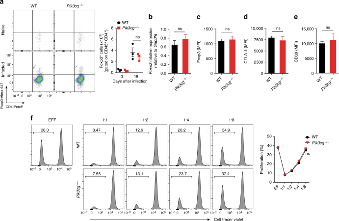
Fig. 5.
The absence of PI3Kγ does not affect the number and suppressive function of regulatory T-cells in the heart tissue of infected mice. a Representative flow cytometry plots for the analysis of Foxp3 staining and quantification of the number of positive cells in the heart tissue of WT (n = 4) and Pik3cg−/− (n = 3) naive mice or infected with T. cruzi Y strain. b PCR analysis of mRNA expression of Foxp3 gene in the heart tissue of WT (n = 4) and Pik3cg−/− (n = 4) mice. Gapdh was used as a housekeeping gene. c–e Flow cytometry analysis measured by the mean fluorescence intensity (MFI) of the cell surface markers Foxp3 (c), CTLA-4 (d), and CD39 (e) expressed in CD4+Foxp3+ cells from the heart tissue of infected of WT (n = 4) and Pik3cg−/− (n = 4) mice 18 days after infection with 103 trypomastigote forms of T. cruzi Y strain. f In vitro T-cell suppression by Treg cells isolated from the lymph nodes of WT and Pik3cg−/− naive mice. ns = no statistical significance (unpaired Student’s t-test in a–f). Data are from one experiment (f) or representative of two (a–e) independent experiments (mean ± s.e.m in a–f)

