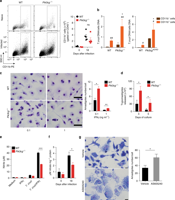
Fig. 7.
Disruption of PI3Kγ signaling in human or murine macrophages harms the killing of intracellular T. cruzi. a Representative flow cytometry dot plots for the analysis of CD11b staining and quantification of the absolute number of positive cells in the heart tissue of non-infected WT (n = 5) and Pik3cg−/− (n = 5) mice or 18 days post infection with 103 trypomastigote forms of T. cruzi Y strain. b Quantitative PCR analysis of nanogram of T. cruzi DNA presents in 1 ng of DNA from CD11b− and CD11b+ cells isolated from the heart tissue of WT and Pik3cg−/− or Pik3cgKD/KD-infected mice. c Representative images of BMDMs stimulated with 0.1 or 1 ng of IFN-γ and infected for 48 h with trypomastigote forms of T. cruzi Y strain at MOI 3:1. Scale bars = 100 µm. d Trypomastigote forms of T. cruzi Y strain released from culture of WT and Pik3cg−/− BMDM stimulated with 0.1 ng ml− of IFN-γ. Experiments were performed in triplicate. e Nitrite quantification by Griess reaction in the supernatant of WT and Pik3cg−/− BMDMs stimulated with 1 ng of IFN-γ and infected with T. cruzi Y strain at MOI 3:1. f Nitrate quantification in the heart tissue of WT (n = 7) and Pik3cg−/− (n = 7) naive mice or 18 days post infection with 103 trypomastigote forms of T. cruzi Y strain. g Representative images of human macrophage lineage THP-1 cells treated with specific PI3Kγ inhibitor AS605240 (1 µM) and infected with trypomastigote forms of T. cruzi Y strain at MOI 3:1. Scale bars = 20 µm. ns = no statistical significance (unpaired Student’s t-test in a). *P < 0.05 relative to WT CD11b+ cells and #P < 0.05 and ##P < 0.01 relative to WT or Pik3cg−/− CD11b− cells (unpaired Student’s t-test in b). *P < 0.05, **P < 0.01, and ***P < 0.001 (unpaired Student’s t-test c–g). Data are one experiment (g) or representative of two (a–d, f) or three (e) independent experiments with similar results (mean ± s.e.m in a–g)

