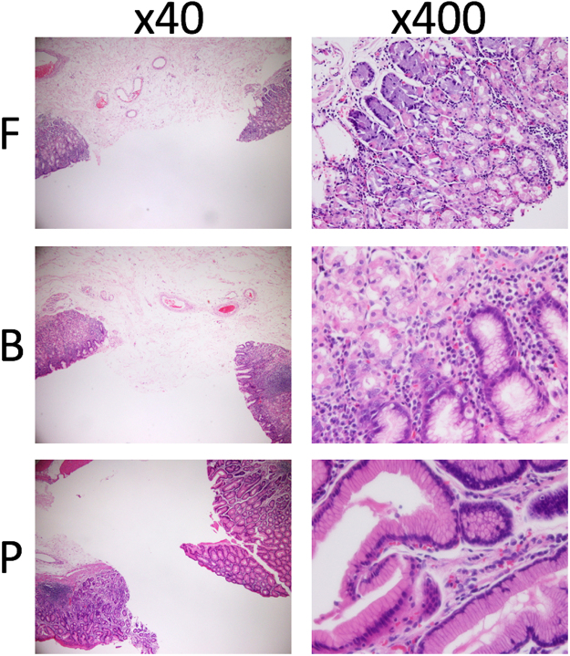
Figure 3.
Haematoxylin and eosin staining to assess gastric glands. All non-tumour areas including the sampled area were subjected to haematoxylin and eosin staining. The adjacent sampled gland area was distinguished as the pyloric gland (P), fundic gland (F), or their combination (borderline; B). When both ends were all P or F glands, the sampled area was considered as P or F. When one end was observed as a combination of P and F glands, the sampled area was considered as B. If one end was P and the other was F, the sampled area was considered as B.

