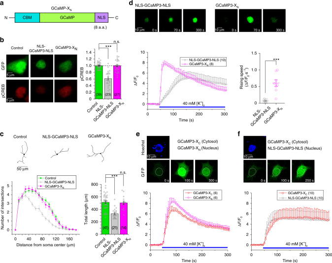
Fig. 7.
Characterizations and validations of GCaMP-XN targeting nuclear Ca2+. a Design of GCaMP-XN. Based on the design principle of GCaMP-X, similar to GCaMP-XC, CBM was fused into N-terminus of GCaMP; and a nuclear localization signal (NLS) was tagged onto C-terminus of GCaMP. b, c Basic validations of GCaMP3-XN with pCREB signals and neurite outgrowth. Representative images of pCREB immunostaining (b, left), statistical summary of pCREB intensities (b, right), tracing of neurite morphology (c, upper), Sholl analysis and statistical summary of neurite length (c, lower) were compared among neurons transfected with YFP, NLS-GCaMP3-NLS or GCaMP3-XN. d Different Ca2+ dynamics resulted from neurons expressing NLS-GCaMP3-NLS or GCaMP3-XN. Confocal images representing Ca2+ fluorescence at three phases: before, during and at the end of extracellular stimuli of 40 mM [K+]o (upper). Ca2+ response (ΔF/F0) (lower left) and its rising speed (ΔF/F0·s−1, normalized change of fluorescence per second, lower right) were averaged from multiple neurons (number indicated within parentheses), to compare NLS-GCaMP3-NLS with GCaMP3-XN. e, f Simultaneous monitoring of cytosolic and nuclear Ca2+ dynamics. Representative confocal images indicative of Ca2+ fluorescence (upper in green) and time-dependent responses (ΔF/F0, lower) are to compare Ca2+ dynamics in the cytosol vs. the nucleus of the same neuron upon 40 mM [K+]o stimuli. Cytosolic Ca2+ was monitored by GCaMP3-XC for both cases, whereas nuclear Ca2+ was either by GCaMP3-XN (e) or by NLS-GCaMP3-NLS (f). Neurons were loaded with Hoechst 33342 to label the nuclei of neurons (upper, blue). Standard error of the mean (S.E.M.) and Student’s t-test (two-tailed unpaired with criteria of significance: *p < 0.05; **p < 0.01, and ***p < 0.001) were calculated when applicable, and n.s. denotes “not significant”

