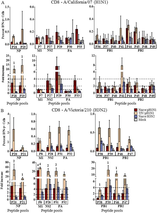
Figure 5.
CD8+ T cell responses to influenza peptide pools. Splenocytes were collected from naïve ferrets infected intranasally with A/NY/21/2009 (Naïve/H1N1pdm09, n = 4), A/Perth/16/2009 (Naïve/H3N2, n = 4), or mock infected ferrets (Naïve/Mock, n = 4) 14d post infection. Splenocytes were also collected from ferrets vaccinated with commercial 2011–2012 TIV, challenged with A/NY/21/2009 (TIV/H1N1pdm09, n = 4). Splenocytes were stimulated with peptide pools (p) derived from regions of each influenza protein from A/California/07/2009 (H1N1pdm09; (A) or the A/Perth/16/2009-like seasonal H3N2 strain, A/Victoria/210/2009 (B). T cells producing IFN-γ in response to peptide stimulation were assessed by flow cytometry. The percent IFN-γ + cells and fold-increase over mock infected animals are depicted. Responses significantly 1greater (p < 0.05) than Mock infected ferrets were considered modest responses, 22-fold greater were considered moderate, and 44-fold greater were considered substantial. Dotted lines indicate 1-, 2-, and 4-fold increases over mock infected controls. Error bars represent SEM.

