Figure 1.

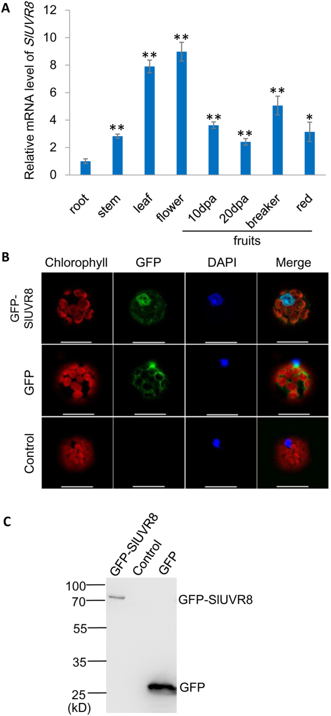
The expression pattern and sub-cellular localization of SlUVR8. (A) Constitutive expression of tomato UVR8 in various tissues. The mRNA levels for tomato UVR8 was analyzed by quantitative RT-PCR. Total RNAs were extracted from roots, stems, leaves, flowers and fruit pericarps at various developmental stages (10, and 20 days post-anthesis, breaker and ripe, respectively). The roots were harvested from plant grown indoor (22–28 °C, 16 h light and 8 h dark) and the rest tissues were harvested from plant grown in the outdoor field. Each bar represents mean value from three biological replicates from each type of tissues (n = 3). Error bars representing standard deviations (SD) are shown in each case. “*” and “**” means P < 0.05 and P < 0.001 respectively (Student’s t test). (B) Localization of GFP-UVR8 fusion protein transiently expressed in tobacco protoplasts. Upper panels, GFP-UVR8; middle panels, GFP; bottom panels, an untransformed protoplast as a negative control. Left to right: red, chlorophyll autofluorescence; green, GFP fluorescence; blue, nucleus stained with DAPI; merged, combined fluorescence from GFP, chlorophyll and DAPI. Scale bars = 25 μm. (C) Western-blot analysis of transient expression samples from (B) by using anti-GFP antibody. The positions of protein ladders were marked on the left side of the gel figure.
