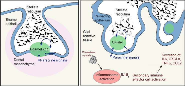
Fig. 10.
Schematic summary of the findings. Molecular and histological relationships between ACP pathogenesis and tooth development. The enamel knot and the β-catenin-accumulating clusters, which both have similar expression profiles and comparable histology, act as signalling hubs through the secretion of a several growth factors acting in an autocrine and/or paracrine manner on the surrounding cells, i.e. the enamel epithelium/dental mesenchyme in the forming tooth or the palisading epithelium, stellate reticulum and glial reactive tissue in ACP. Reciprocal signalling from surrounding tissues to the enamel knot and clusters is indicated by double-headed arrows. In the glial reactive tissue, cholesterol crystals activate the inflammasomes resulting in the secretion of IL1B, which in turn acts on the local immune effector cells to drive an inflammatory response

