Fig. 2.
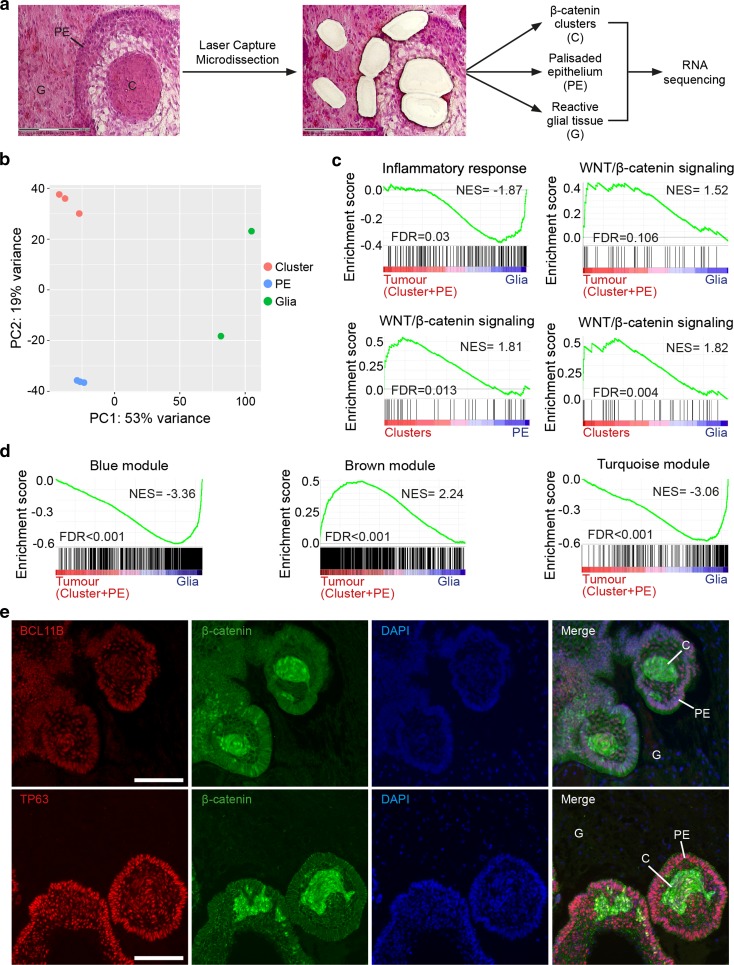
Gene profiling of laser capture microdissected tumour cells confirms the WGCNA, identifying modules associated with tumour cell compartments and revealing novel ACP genes. a Scheme of the experimental approach. Histological sections of tumour samples JA004 and JA029 were subjected to laser capture microdissection (LCM) to isolate β-catenin-accumulating cell clusters (C), palisading epithelium (PE) and glial reactive tissue (G). Biological duplicates were performed for clusters and palisading epithelium in case JA004. RNA was purified from each of these tumour cell compartments, amplified and sequenced. b Principal component analysis reveals grouping of the data from laser capture microdissected samples. c Gene set enrichment analysis revealing the enrichment of an inflammatory signature in microdissected glial reactive tissue relative to tumour tissue (i.e. genes included in clusters plus palisading epithelium), whilst a WNT signalling expression signature is associated with the microdissected tumour tissue. Enrichment for WNT signalling was stronger in the clusters relative to both PE and glial tissue. d Gene set enrichment analysis showing the enrichment of the brown module genes with a signature of tumour cell compartments (i.e. genes including in cluster cells plus palisading epithelium). In contrast, both the blue and turquoise module genes are predominantly expressed by reactive glia. e Double immunofluorescence staining revealing the expression of BCL11B and TP63 in the epithelial components of the tumour, including palisading epithelium (PE) and β-catenin accumulating clusters (C), but not in reactive glial tissue (G). NES normalised enrichment score, FDR false discovery rate. Scale bars 100 μm

