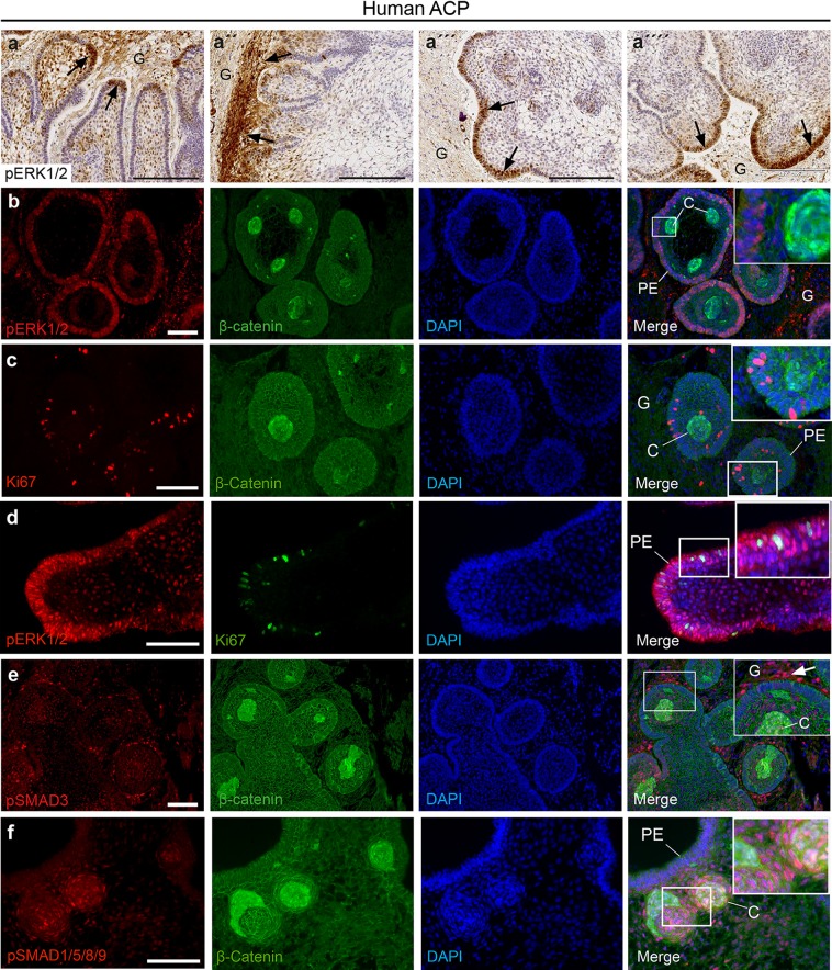
Fig. 4.
Identification of the activation of the MAPK/ERK, TGFB and BMP signalling pathways in human ACP. a Immunohistochemistry revealing the expression of phosphorylated ERK1/2 (pERK1/2), a read out of active MAPK/ERK pathway, at the tips of the invading tumour epithelium (palisading epithelium, arrows in a, a‴ and a″″) and within reactive glial tissue (G; arrows in a″). b Double immunofluorescent staining showing pERK1/2 expression in the palisading epithelium (PE) around the β-catenin accumulating clusters (C), which express several activating ligands of the MAPK/ERK pathway (see main text for details). Note that cells within the reactive glial tissue (G) are also pERK1/2 positive. c Double immunofluorescence revealing abundant Ki67+ve cells in the palisading epithelium close to clusters. d Double immunofluorescence showing Ki67 and pERK1/2 co-expression within the palisading epithelium (PE). e Double immunofluorescence showing pSMAD3 staining, indicating activation of TGFβ signalling, in both tumour and reactive glia, with strongest signal in reactive tissue adjacent to tumour epithelia (arrowhead). Double immunofluorescence reveals pSMAD1/5/9 staining, indicating BMP signalling in cells within and adjacent to the β-catenin-accumulating clusters (C). Note the absence of staining in the palisading epithelium (PE). Scale bars: a–a″″ 200 μm; b–f 100 μm

