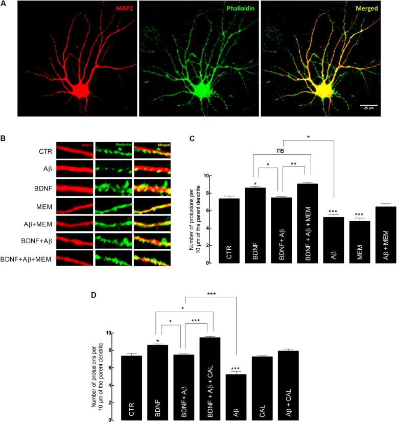
FIGURE 3.
Brain-derived neurotrophic factor (BDNF) restores its capacity to increase spine density after inhibition of Aβ-induced NMDAR activation. (A) Representative image of an untreated neuron obtained from primary cultures. DIV14 neurons were incubated with BDNF (20 ng/mL) for 24 h, in the presence or absence of Aβ25–35 (25 μM) and/or memantine (1 μM) or calpastatin (1 μM). MAP2 (red) specifically detects neurons, while phalloidin (green), recognizes F-actin, thus labeling protrusions (filopodia and spines). The merge of both elements is represented in yellow. Six neurons were analyzed per condition and spine density was considered, in each cell, as the number of protrusions per 10 μm of the parent dendrite with a distance of 25 μm from the cell body. Protrusions were counted in each neuron in three different dendrites. (B) Treatments effects on synaptic growth. Aβ significantly reduces the number of protrusions, whereas BDNF increases the number of protrusions when incubated alone. In the presence of Aβ, BDNF loses its ability to increase the number of protrusions, which is rescued when cells are incubated with memantine. (C) The panels show the average number of protrusions in different conditions when neurons were treated with memantine and (D) calpastatin (∗p < 0.05, ∗∗p < 0.01, ∗∗∗p < 0.001, n = 6–14, three-way ANOVA with Tukey post hoc test). Values are mean ± SEM.

