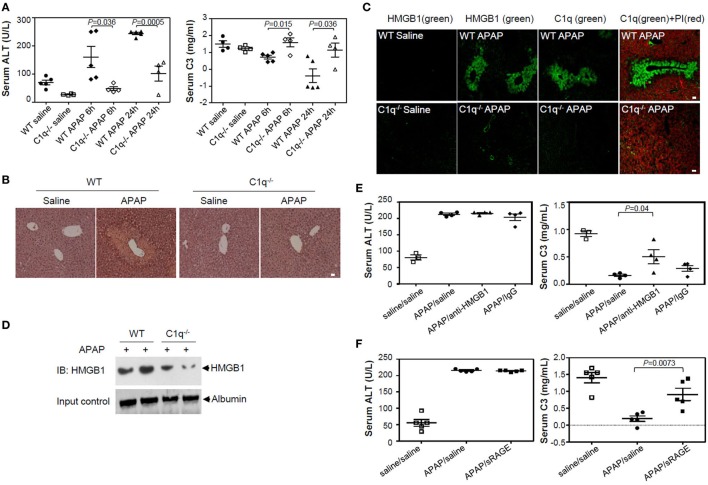
Figure 6.
High-mobility group box 1 (HMGB1)-induced complement activation is decreased in C1q-deficient mice of APAP-induced hepatotoxicity model. (A) Overnight fasted wild-type (WT) or C1q−/− mice (9–10 weeks old, male) were intraperitoneally (i.p.) treated with 400 mg/kg of APAP or saline. After 0, 6, and 24 h, mouse serum samples were collected. Serum alanine aminotransferase (ALT) activity (left) and C3 levels (right) were determined using ALT color endpoint assay and enzyme-linked immunosorbent assay, respectively. Each dot represents each mouse with triplicates per sample (four or five mice per group). (B,C) Mice were euthanized 24 h after APAP administration, and the left medial lobe of liver was fixed in 4% paraformaldehyde and 30% sucrose. Liver tissue was stained with hematoxylin and eosin for evaluation of necrosis and hemorrhage (B). Tissue-Tec OCT-embedded liver was stained with anti-C1q and anti-HMGB1 antibodies (Abs) for immunofluorescence analysis (C). (D) Serum HMGB1 protein from WT and C1q−/− mice was detected using Western blot analysis 24 h after APAP injection. (E) Anti-HMGB1 (2G7) or mouse IgG (5 μg/mouse) was i.p. administrated immediately after APAP administration to block HMGB1. After 24 h, mouse serum samples were collected and ALT activity (left panel) and concentration of C3 (right panel) were determined. Saline was used as a negative control. (F) HMGB1 was i.p. injected with sRAGE (5 µg/mouse) as described in (E). N = 4–5 mice/group. Data = mean ± SEM. Student’s t-test (unpaired two-tailed) was used to calculate the P-value. All scale bars, 20 µm. Data are representative of three (A–E) or two (F) independent experiments.

