Figure 5.

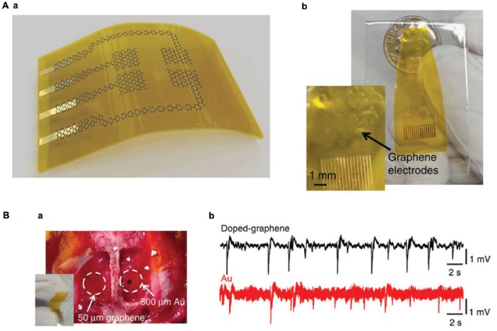
Graphene electrodes for in vivo recording. (A) (a) Schematic illustration of a flexible G neural electrode array. (b) Photograph of a 16-electrode transparent array. The electrode size is 300 × 300 μm2. (B) (a) Photograph of a 50 × 50 μm2 single-G electrode placed on the cortical surface of the left hemisphere and a 500 × 500 μm2 single-Au electrode placed on the cortical surface of the right hemisphere. (b) Interictal-like spiking activity recorded by 50 × 50 μm2 doped-G and Au electrodes. Recordings with doped-G electrodes are five- to sixfold less noisy compared with the ones with same size Au electrode. Modified with permission from Kuzum et al. (2014).
