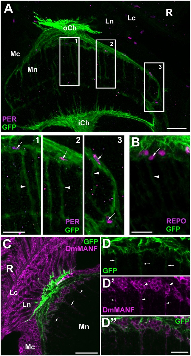
Figure 6.

(A) Horizontal section of the medulla of NP6520-Gal4/UAS-mCD8-GFP flies immunolabeled with anti-PER antibody. The nuclei of GFP-expressing EnGl (frames 1–3) are PER-positive, which is well visible in higher magnification (panels 1–3). The processes of EnGl (arrowheads in panels 1–3) span the medula neuropil (Mn). R, retina; Lc, Lamina cortex; Ln, lamina neuropil; oCh, outer chiasm; Mc, medulla cortex; iCh, inner chiasm. Scale bars: 20 μm for (A) and 10 μm for panel 1, 2, and 3. (B) The nuclei of GFP-positive EnGl show high level of REPO-specific immunofluorescence (arrow). Arrowhead-the EnGl processes. Scale bar: 10 μm. (C) The distal part of the medulla in horizontal section of NP6520-Gal4/UAS-mCD8-GFP flies immunolabeled with anti-DmMANF antibody. The processes of EnGl are marked with arrows. R, retina; Lc, lamina cortex; Ln, lamina neuropil; Mc, medulla cortex; Mn, medulla neuropil. Scale bar: 20 μm. (D-D”) Higher magnification of EnGl marked in (C). The GFP-positive processes of EnGl (D, arrows) are marked with DmMANF (D',D”). DmMANF is also visible in the perinuclear space of cell bodies in the medulla cortex (D', arrowheads). Scale bar: 10 μm.
