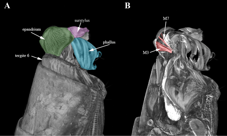
Figure 1.
Micro-CT surface rendering (A) and volume rendering of virtual sections to median, digitally stained (B) of Nothybus kuznetsovorum (Nothybidae), lateral view. Cerci shown in yellow, epandrium in dark green, phallus in light blue, surstylus in pink, syntergosternite VIII in violet, syntergosternite VII in light green, sternite VI in orange and sternite V in dark blue.

