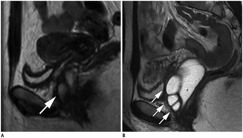
Fig. 12. 49-year-old woman complaining of mass in lower part of vagina.
A. Vaginal abnormalities are observed in sagittal T2-weighted MR images without vaginal opacification, but are difficult to analyze (arrow). B. After vaginal opacification was conducted subsequently, three cysts of anterior vaginal wall became much more conspicuous (arrows).

