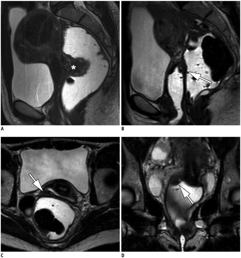
Fig. 3. Pre-operative MR images in 37-year-old patient with known endometriosis and suffering from recurrent dyschesia.
Sagittal T2-weighted MR view with rectal opacification (A) revealing endometriotic nodule of anterior wall of rectum (*), surgically removed. MRI conducted after surgery using T2-weighted MR images after vaginal and rectal opacification in sagittal (B), axial (C), and coronal (D) planes demonstrates endometriotic nodule in lateral right vaginal fornix (arrows) (B-D). This lesion had not been detected on pre-operative MR imaging, probably because of absence of initial vaginal distension.

