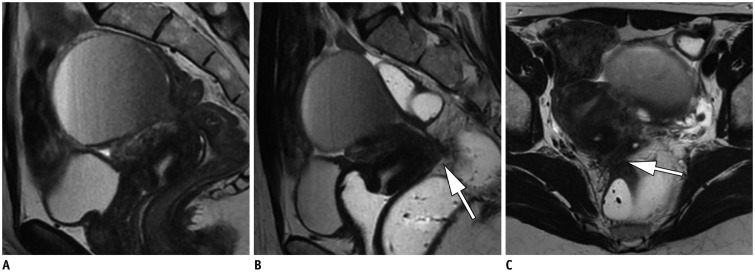
Fig. 4. Pre-operative pelvic MR study in 28-year-old female with endometriosis presenting pain radiating to back.
Sagittal T2-weighted MR image using vaginal and rectal opacification reveals thick lesion in posterior vaginal fornix and posterior cervix (arrow) (B). There is large nodular lesion infiltrating anterior rectal wall on axial T2-weighted MR image (arrow) (C), associated with Douglas cul-de-sac obliteration. Vaginal and rectal opacification clearly improve detection of these lesions compared to what was observed on MR image MRI study conducted without opacification (A).

