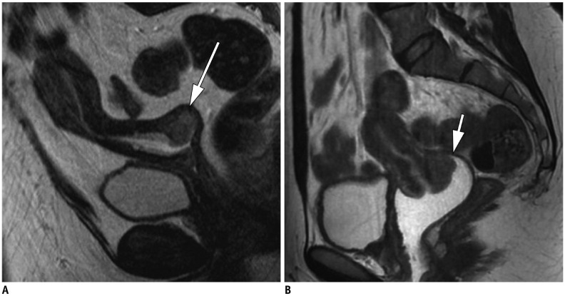
Fig. 5. Sagittal T2-weighted MR images without and after vaginal opacification in two women (respectively 41-year-old and 47-year-old) with cervical carcinoma.
Relatively high signal-intensity tumor is observed in posterior cervix (arrow). Vaginal fornices appear to be invaded by mass in absence of vaginal distension (A). After vaginal opacification, borders of tumor are better observed, and no vaginal extension is depicted in second case (arrow) (B). Tumor′s boundaries are less conspicuous without opacification.

