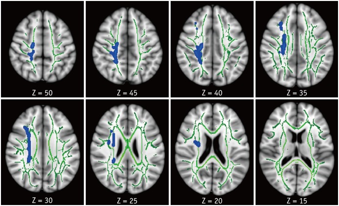
Fig. 1. TBSS analysis of FA maps.
Areas in sky blue-blue represent brain regions with significant decrease in FA (FWE-corrected p < 0.05) in HAND group relative to HC. Results are shown overlaid on Montreal Neurological Institute 152-T1 template and mean FA skeleton (green). Left side of image corresponds to right hemisphere of brain. FA = fractional anisotropy, FWE = family-wise error, HAND = HIV-associated neurocognitive disorders, HC = healthy controls, HIV = human immunodeficiency virus, TBSS = tract-based spatial statistics

