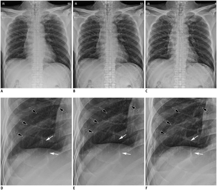
Fig. 1. Comparison of (A, D) non-grid, (B, E) grid-like, and (C, F) grid images.
A-C. Chest radiography of 57-year-old male patient who underwent multiple-wedge resection in both lungs for metastasis associated with undifferentiated pleomorphic sarcoma. Note superior image contrast of grid-like image to non-grid image, and similarity in contrast appearance between grid-like and grid images. D-F. Magnified non-grid, grid-like and grid images: surgical materials (black arrows) and multiple linear opacities (white arrows) are clearly demonstrated in grid-like image, as well as grid image.

