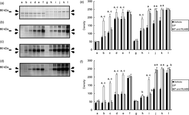
Figure 3.

Acceleration and increasing of tyrosine phosphorylation of 80‐kDa sperm proteins by progesterone (P). (a) Typical Coomassie Brilliant Blue‐stained membrane after blotting. (b) Western blotting against proteins obtained from spermatozoa that were incubated in vehicle. (c) Western blotting against proteins obtained from spermatozoa that were incubated in 20 ng/mL P (P). (d) Western blotting against proteins obtained from spermatozoa that were incubated in 20 ng/mL P and RU486 (P and RU486). Arrows show tyrosine phosphorylation of 80‐kDa sperm proteins. Numbers on the left‐hand side indicate the molecular weight standard of 80 kDa. (e) Density of upper bands detected on (b–d). (F) Density of lower bands detected on (b–d). aSignificantly different than vehicle (P < 0.01); bsignificantly different than vehicle (P < 0.05); csignificantly different than P and RU486 (P < 0.01). Lanes a–f and g–l illustrate the results from urea extract and urea–thiourea extract, respectively. Lanes a and g were incubated for 0 h after supplying P. Lanes b and h were incubated for 0.5 h, lanes c and i were incubated for 1 h, lanes d and j were incubated for 2 h, lanes e and k were incubated for 3 h, and lanes f and l were incubated for 4 h.
