
Figure 2.
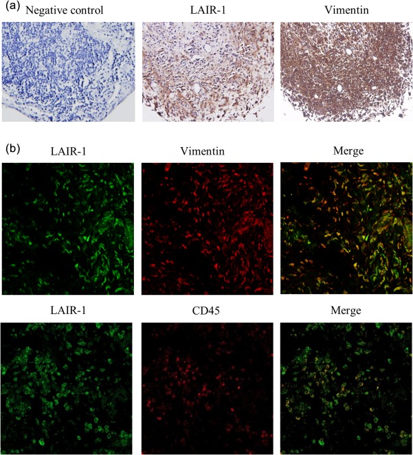
Immunolabelling of leucocyte‐associated immunoglobulin (Ig)‐like receptor‐1 (LAIR‐1) and vimentin in human rheumatoid arthritis (RA) synovial tissue. (a) Consecutive synovial sections from the RA patients were used to detect LAIR‐1 and vimentin. Levels of LAIR‐1 expression were higher in the synovial lining than the sublining layer. IgG isotype served as the negative control in the staining; ×400 magnification. (b) Co‐localization of LAIR‐1/vimentin and LAIR‐1/CD45 in the synovial lining was detected by immunofluorescence staining and confocal microscopy; ×400 magnification. These results were confirmed by examining synovial membrane specimens from six RA patients. Immunolabelling of a representative sample is shown.