
Figure 3.
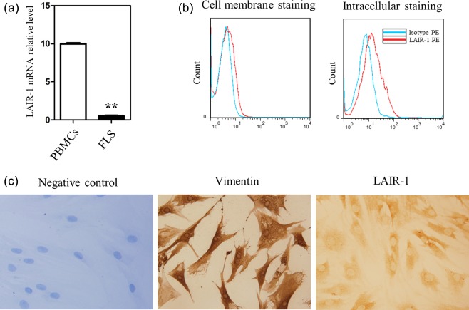
Expression of leucocyte‐associated immunoglobulin (Ig)‐like receptor‐1 (LAIR‐1) in primary fibroblast‐like synoviocytes (FLS) isolated from the rheumatoid arthritis (RA) patients. (a) Levels of LAIR‐1 mRNA in normal human peripheral peripheral blood mononuclear cells (PBMC) (n = 3) and FLS (n = 5) as shown by quantitative polymerase chain reaction (qPCR). Reaction without template cDNA served as the negative control, and showed no melting curve. **P < 0·01 versus the PBMC group. (b) Flow cytometric analysis of the LAIR‐1‐positive FLS populations from the RA patients (n = 11). Intracellular staining and flow cytometric analysis of LAIR‐1 were also performed with phycoerythrin (PE)‐labelled monoclonal antibodies (mAbs) (n = 5). Representative flow cytometry graphs are shown. (c) Immunohistochemical images show the expression of LAIR‐1 and vimentin in the primary FLS isolated from the RA patients. IgG isotype‐stained sections did not stain positively; ×400 magnification. Data are representative of FLS from six different RA patients.