
Figure 4.
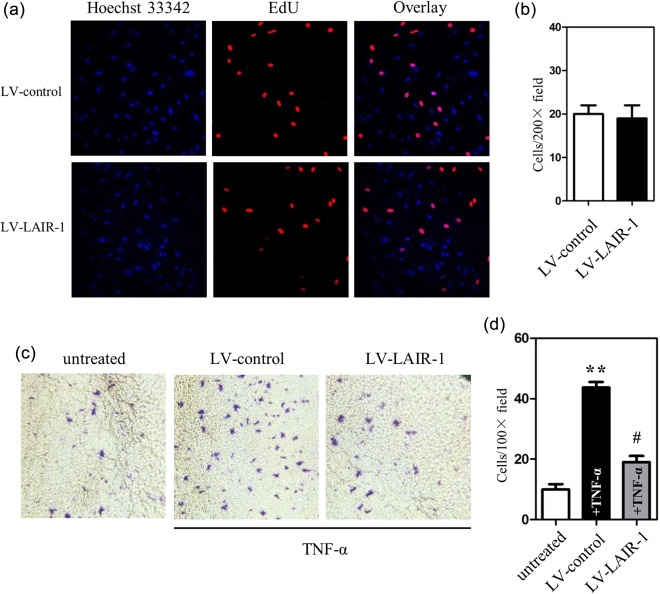
Role of leucocyte‐associated immunoglobulin (Ig)‐like receptor‐1 (LAIR‐1) in the modulation of the cell proliferation and invasion of fibroblast‐like synoviocytes (FLS). (a) MH7A cells at a density of 5 × 10³ cells per well were cultured in 96‐well plates and transfected with either a negative control lentiviral vector (LV‐control) or the LAIR‐1 overexpression lentiviral vector (LV‐LAIR‐1) for 48 h. Cells were then exposed to 5‐ethynyl‐2'‐deoxyuridine (EdU) (50 μM) for 4 h, followed by staining with the Apollo reaction cocktail for 30 min. EdU‐labelled proliferating cells were examined by fluorescence microscopy. Data shown are representative images of individual groups (n = 6 per group) from three independent experiments; ×200 magnification. (b) Cell numbers per microscopic field were plotted in the histogram. (c) LV‐LAIR‐1 was used to study the role of LAIR‐1 in tumour necrosis factor (TNF)‐α‐induced FLS invasion. Photomicrographs show cells that have passed through the membrane. MH7A cells at a density of 4 × 10⁴ cells per well were cultured in a 24‐well Transwell insert. After infection with LV‐LAIR‐1 or LV‐control for 24 h, cells were treated with TNF‐α. FLS were allowed to invade for 24 h; ×200 magnification. (d) Cell number per microscopic field was plotted in the histogram. **P < 0·01 versus the untreated control group. #P < 0·05 versus TNF‐α‐treated LV control cells.