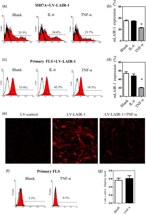
Figure 5.

Tumour necrosis factor (TNF) decreased leucocyte‐associated immunoglobulin (Ig)‐like receptor‐1 (LAIR‐1) expression significantly at the cell surface. (a,b) Flow cytometric analysis and statistics of the MH7A cells infected with lentiviral vector (LV)‐LAIR‐1 for 24 h. MH7A cells were then treated with IL‐6 or TNF‐α (10 ng/ml) for another 24 h. A blank group served as the control. *P < 0·05 versus the blank group and data were obtained from three independent experiments. (c,d) Flow cytometric analysis and statistics of primary fibroblast‐like synoviocytes (FLS) from rheumatoid arthritis (RA) patients infected with LV‐LAIR‐1 (n = 3). FLS were treated with the same conditions as the MH7A cells. (e) Images obtained by laser scanning confocal microscopy showing LAIR‐1 expression (red). MH7A cells were infected with the LV‐control or LV‐LAIR‐1 lentiviral vector. LV‐LAIR‐1‐infected cells were then stimulated in the presence or absence of TNF‐α (10 ng/ml) for 24 h; ×100 magnification. (f) Flow cytometric analysis of cell‐surface LAIR‐1 expression in primary FLS from RA patients (n = 3). Cells were treated with TNF‐α (10 ng/ml) for 24 h, and a blank group was included as the control. (g) Primary FLS from RA patients treated with TNF‐α for 24 h were collected, and then RNA was extracted for qPCR analysis, and a blank group was included as the control (n = 3).
