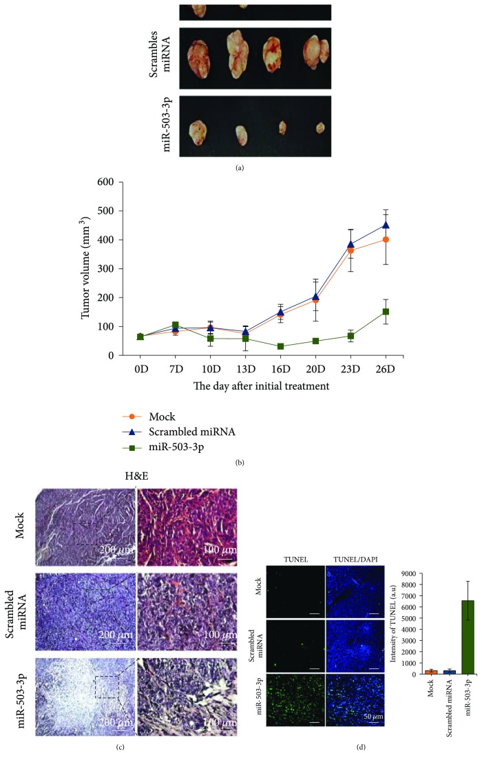
Figure 4.
miR-503-3p exerts antitumor activity on MCF7 xenografts. (a) The dissected tumors are shown at 28 days postinitial treatment. (b) Tumor growth curves after initial treatment. miRNA-NC and miR-503-3p were intratumorally administered every 3 days for 2 weeks, and tumor volumes were monitored every 3 days. Error bars indicate SDs (n = 4). (c) Histologic observation was conducted using hematoxylin and eosin (H&E) staining. Right panels indicate the magnified images from the dotted-line boxes. (d) TUNEL assay was used to detect apoptotic cells in tissues. Apoptotic cells were visualized by confocal microscopy and quantified using ImageJ.

