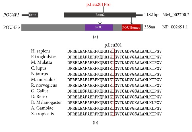
Figure 2.
Conservation analysis and genomic structure of POU4F3 based on the open reading frame (NM_002700.2) containing 2 exons (black rectangles). (a) The position of POU4F3 c.602T>C (p.Leu201Pro) is highlighted in red and shown both at the gene (top) and the protein level (bottom). The protein diagram depicts the predicted functional domains and sequence motifs. (b) Protein alignment showing POU4F3 p.Leu201 occurred at evolutionarily conserved amino acids (in red box) across twelve species.

