Figure 2.
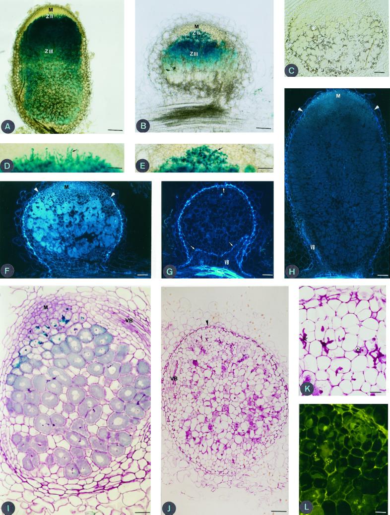
Longitudinal thick sections (80 μm) of DZA315.16 (A) and Jemalong 6 (B) nodules 20 DPI induced by S. meliloti strain A145 (pXGLD4), showing typical zonation of an effective M. truncatula nodule, with a meristematic zone (M), infection zone (ZII), and the nitrogen-fixing zone (ZIII). Bar = 100 μm. Section (4 μm thick) of a Technovit-embedded nodule of Jemalong 6 (C) 20 DPI after staining with iodine. Starch accumulation (asterisks) is observed in the whole nodule and not solely localized to an amyloplast-rich interzone II-III layer. Bar = 10 μm. Longitudinal sections (80 μm thick) of DZA315.16 (D) and Jemalong 6 (E) nodules 20 DPI. Note that DZA315.16 infection threads are thin and elongated, those of Jemalong 6 are short and hypertrophied. Infection threads are indicated by black arrows. Bar = 50 μm. Longitudinal and median thin sections (4 μm) of Jemalong 6 nodule 20 DPI (F) and 30 DPI (G) under UV illumination. Note the absence of meristematic zone and the autofluorescence of endodermal cells around the whole nodule and the vascular bundles (VB) in G compared to F. White arrows indicate endodermal cells at the distal part of nodule and around vascular bundles. Bar = 10 μm. Longitudinal and median thin section of DZA315.16 nodule 30 DPI (H); white arrowheads indicate the limit of endodermal cells. Bar = 10 μm. Thin section of Jemalong 6 nodule, viewed under light microscopy after staining with toluidine blue (I), at 20 DPI dividing meristematic cells (M) are observed. Bar = 50 μm. Thin section of Jemalong 6 nodule 30 DPI (J); note the presence of numerous empty cells indicating senescence of the nodule. Bar = 100 μm. Thin section (4 μm) through 30-d-old nodule of Jemalong 6 viewed under light microscopy (K), infection threads (stars) are thick and short and under blue light (L), note the autofluorescence of infection threads (stars). Bar = 20 μm.

